Article-Journal

All-in-One Molecular Aggregation-Induced Emission Theranostics: Fluorescence Image Guided and Mitochondria Targeted Chemo- and Photodynamic Cancer Cell Ablation
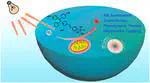
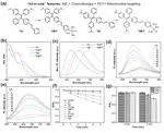
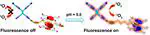
Molecular theranostic platforms with precise molecular structure and multiple functions hold great promise for cancer therapy. Different from the current strategy to incorporate various components into single entities with the risk of compromised efficacy and poor reproducibility, herein, molecular aggregation-induced emission (AIE) photosensitizers with ingenious integration of AIE fluorophore and cisplatin are facilely synthesized for synergetic anticancer therapy. Through adjusting donor structures coordinated with a cisplatin moiety and balancing the hydrophobic–hydrophilic property, donor–acceptor strength, and intramolecular charge transfer effect, the newly designed AIE photosensitizer TNPT exhibits good cellular uptake with predominant mitochondria location of cancerous cells, high chemotherapeutic efficacy similar to that of cisplatin, and strong reactive oxygen species (ROS) generation capability better than that of chlorin e6 (Ce6). Importantly, TNPT demonstrates synergetic photodynamic and chemotherapy on C6 glioma cells, showing 2.4-fold more potency than cisplatin upon white light irradiation (15 J/cm2). Further cell cycle analysis and apoptosis assay indicate that the photodynamic and chemo-therapeutic functions of TNPT synergistically inhibit DNA replication and cause cell apoptosis. In addition, TNPT exhibits selective uptake on cancerous cells rather than normal cells, contributing to significantly lower cytotoxicity to normal cells as compared to free cisplatin. This study provides a facile strategy to design molecular theranostic agents.
All-in-One Molecular Aggregation-Induced Emission Theranostics: Fluorescence Image Guided and Mitochondria Targeted Chemo- and Photodynamic Cancer Cell Ablation
Molecular theranostic platforms with precise molecular structure and multiple functions hold great promise for cancer therapy. Different from current strategy to incorporate various components into single entities with the risk of compromised efficacy and poor reproducibility, herein, molecular aggregation-induced-emission (AIE) photosensitizers with ingenious integration of AIE fluorophore and cisplatin are facilely synthesized for synergetic anticancer therapy. Through adjusting donor structures coordinated with cisplatin moiety and balancing the hydrophobic-hydrophilic prop-erty, donor-acceptor strength, and intramolecular charge transfer effect, the newly designed AIE photosensitizer TNPT exhibits good cellular uptake with predominant mitochondria location of cancerous cells, high chemotherapeutic efficacy similar to cisplatin, and strong reactive oxygen species (ROS) generation capability better than chlorin e6 (Ce6). Im-portantly, TNPT demonstrates synergetic photodynamic and chemo-therapy on C6 glioma cells, showing 2.4-fold more potent than cisplatin upon white light irradiation (15 J/cm2). Further cell cycle analysis and apoptosis assay indicate that the photodynamic and chemo-therapeutic functions of TNPT synergistically inhibit DNA replication and cause cell apop-tosis. In addition, TNPT exhibits selective uptake on cancerous cells rather than normal cells, contributing to significantly lower cytotoxicity to normal cells as compared to free cisplatin. This study provides a facile strategy to design molecular theranostic agents.
All-in-One Molecular Aggregation-Induced Emission Theranostics: Fluorescence Image Guided and Mitochondria Targeted Chemo- and Photodynamic Cancer Cell Ablation
Nanostructural Control Enables Optimized Photoacoustic–Fluorescence–Magnetic Resonance Multimodal Imaging and Photothermal Therapy of Brain Tumor
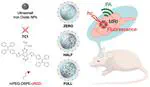
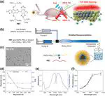
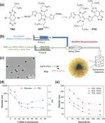
The performance of current multimodal imaging contrast agents is often constrained by the tunability of nanomaterial structural design. Herein, the influence of nanostructure on the overall imaging performance of a composite nanomaterial for multimodal imaging of brain tumors is studied. Newly designed near-infrared molecules (TC1) are encapsulated into nanocomposites with ultrasmall iron oxide nanoparticles (UIONPs), forming stable nanoagents for multimodal imaging and photothermal therapy (PTT). Through a modified nanoprecipitation method, the synthesis of nanocomposites denoted as HALF is realized, in which UIONPs are restricted to half of the nanosphere. Such a unique nanostructure that physically separates TC1 and UIONPs is found with capabilities of mitigating fluorescence quenching, preserving the good performance of photoacoustic imaging, and enhancing the magnetic resonance imaging signals. Decorated with a peptide ligand cRGD for better brain tumor targeting, HALF-cRGD is evaluated both in vitro and in vivo as imaging contrast agents and photothermal therapeutic agents. The good imaging performance and PTT effect of HALF-cRGD in mice models indicate that the rational design and control of nanostructures could optimize multimodal imaging performance using the same components.
Nanostructural Control Enables Optimized Photoacoustic–Fluorescence–Magnetic Resonance Multimodal Imaging and Photothermal Therapy of Brain Tumor
High-Resolution 3D NIR-II Photoacoustic Imaging of Cerebral and Tumor Vasculatures Using Conjugated Polymer Nanoparticles as Contrast Agent
Exogenous contrast-agent-assisted NIR-II optical-resolution photoacoustic microscopy imaging (ORPAMI) holds promise to decipher wide-field 3D biological structures with deep penetration, large signal-to-background ratio (SBR), and high maximum imaging depth to depth resolution ratio. Herein, NIR-II conjugated polymer nanoparticle (CP NP) assisted ORPAMI is reported for pinpointing cerebral and tumor vasculatures. The CP NPs exhibit a large extinction coefficient of 48.1 L g−1 at the absorption maximum of 1161 nm, with an ultrahigh PA sensitivity up to 2 µg mL−1. 3D ORPAMI of wide-field mice ear allows clear visualization of regular vasculatures with a resolution of 19.2 µm and an SBR of 29.3 dB at the maximal imaging depth of 539 µm. The margin of ear tumor composed of torsional dense vessels among surrounding normal regular vessels can be clearly delineated via 3D angiography. In addition, 3D whole-cortex cerebral vasculatures with large imaging area (48 mm2), good resolution (25.4 µm), and high SBR (22.3 dB) at a depth up to 1001 µm are clearly resolved through the intact skull. These results are superior to the recently reported 3D NIR-II fluorescence confocal vascular imaging, which opens up new opportunities for NIR-II CP-NP-assisted ORPAMI in various biomedical applications.
High-Resolution 3D NIR-II Photoacoustic Imaging of Cerebral and Tumor Vasculatures Using Conjugated Polymer Nanoparticles as Contrast Agent